De Winter electrocardiograph ECG pattern signifies proximal left anterior descending coronary artery LAD occlusion and extensive anterior myocardial infarction and it is found in about 2 of patients with proximal LAD occlusion. Rokos I et al.
De Winter T Wave Litfl Ecg Library Diagnosis
Differentiation of this ECG pattern is essential to refer patients for appropriate and immediate reperfusion therapy.

. The pattern was seen in the precordial leads is. In fact the title was Persistent precordial hyperacute T-waves signify proximal left anterior descending artery occlusion. The value of maximal STD culprit artery and culprit site with Thrombolysis In Myocardial Infarction TIMI or Rentrop flow.
In 2008 de Winter et al described an ECG pattern suggesting that it should be considered an ST-elevation myocardial infarction STEMI equivalent de Winter Verouden Wellens Wilde 2008 with the potential to predict critical stenosis or occlusion of the left anterior descending coronary artery LAD. Suggesting acute STEMI without displaying signature ST elevation on the ECG. Earlier percutaneous coronary intervention PCI.
Is observed in about 2 patients with proximal LAD occlusion. A new ECG sign of proximal LAD occlusion. Later described a case of the de Winter ECG pattern evolving to anterior ST-segment elevation consistent with STEMI over a 2 hour period.
An electrocardiographic finding suggestive of impending myocardial infarction the de Winters pattern or de Winters T-waves describes an abnormality thought to be indicative of acute occlusion of the proximal left anterior descending coronary artery LAD 2. We aimed to investigate the morphology of the De Winter ECG pattern and evaluate the test characteristics of the De Winter pattern for the diagnosis of acute coronary occlusion. The de Winter Electrocardiogram Pattern Evolving From Hyperacute T Waves It reminded me that many believe due to the assertions in the original de Winters article that de Winters waves are stable.
Besides the morphology of upsloping or nonupsloping ST depression STD may have different significance of severity and prognostication. Find read and cite all the research you. Complications of acute aortic dissection AD in the setting of acute myocardial infarction AMI with de Winter sign are relatively rare and physicians may easily miss the diagnosis of AD.
The reported positive predictive value PPV for the de Winter ECG pattern to predict an acute left anterior descending artery LAD lesion is inconsistent. The de Winter ECG pattern is a recently-described STEMI equivalent that emergency physicians and paramedics must be aware of. The de Winter ECG pattern indicates occlusion of the LAD and is frequently underrecognized by clinicians which increases morbidity and mortality.
First recognized and reported by de Winter et al. Represents approximately 2 of LAD occlusions. ECG characteristics of De Winters T waves include.
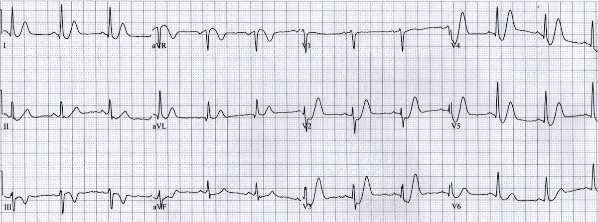
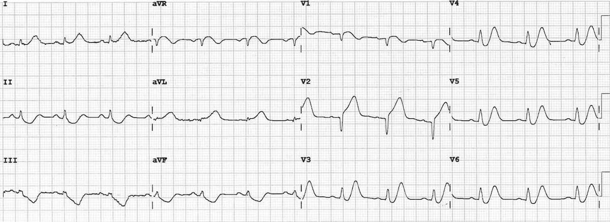
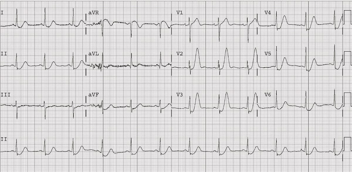
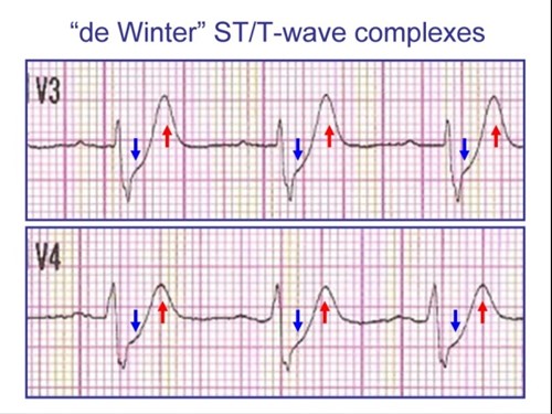
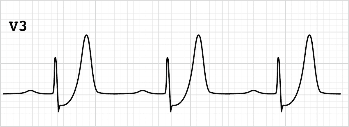
Characterized by 1-3 mm of ST-depression with upright symmetrical T-waves. ST segment depression at the J point of precordial leads of 1-3 mm Up-sloping ST segments becoming peaked positive symmetrical T waves Occurred in leads V1 V6 QRS complexes were in some cases slightly widened Loss of precordial R-wave progression aVR also showed small ST Elevation of 1-2mm. The electrocardiogram ECG is one of the most useful diagnostic studies for identification of acute coronary syndrome ACS and acute myocardial infarction AMI.
ECG abnormality described by de Winter et al. The De Winter ECG pattern has been reported to indicate acute left anterior descending coronary artery occlusion and is often considered to be an ST elevation myocardial infarction STEMI equivalent. First reported by Dutch Professor of Cardiology Robbert J.
De Winter electrocardiograph ECG pattern is an atypical presentation of acute myocardial infarction AMI due to severe stenosis of the left anterior descending LAD. De Winter sign first described by De Winter et al. But Goebel et al.
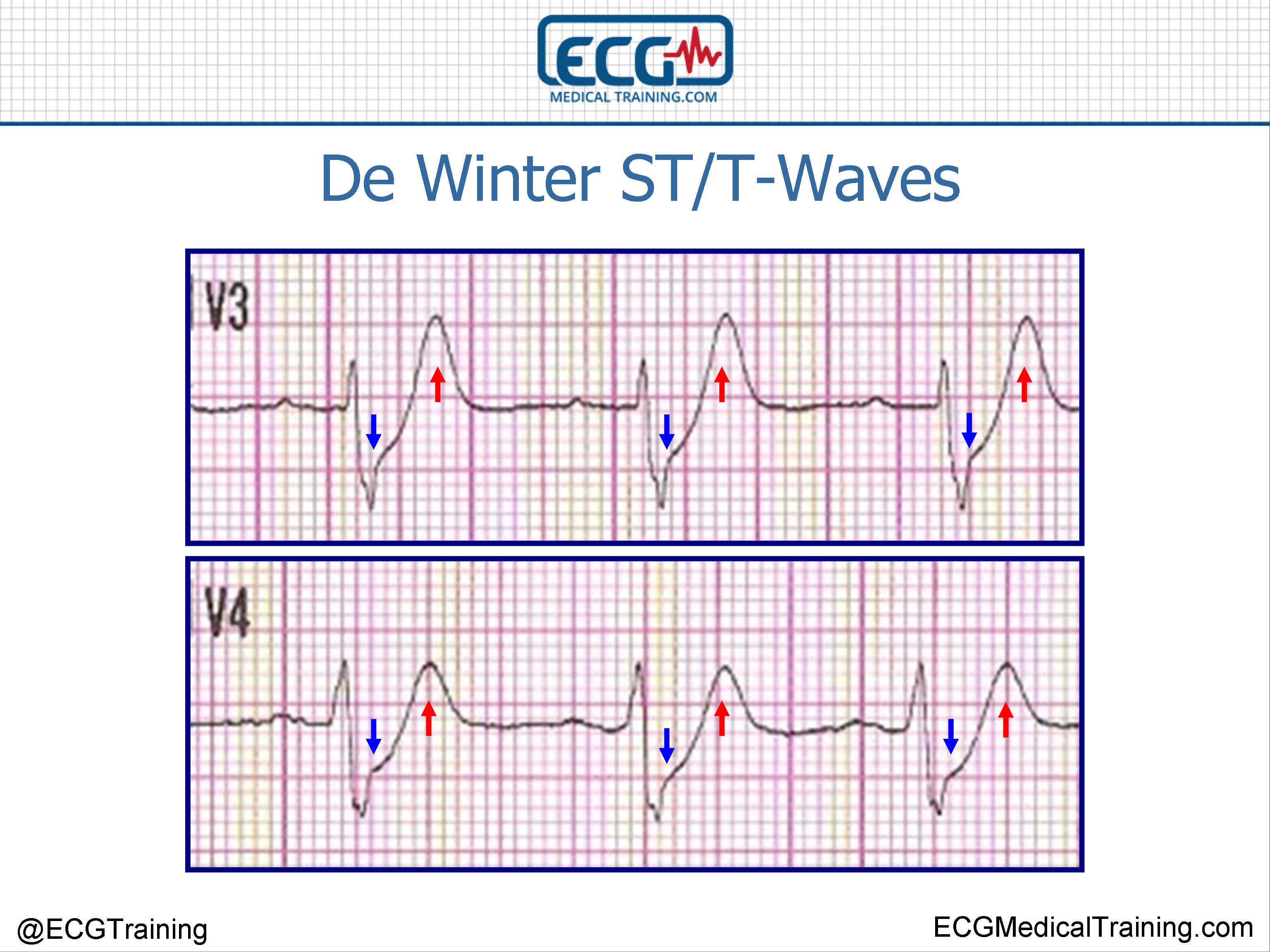
The de Winter ECG pattern was initially described as a static phenomenon 78 ie. Up to 10 cash back The de Winter ECG pattern characterized by 13-mm upsloping ST-segment depression at the J point in leads V1 to V6 that continues to tall positive symmetrical T waves is identified in only 2 of the patients with acute MI 1. Very slight 05-1mm ST elevation in lead aVR.
1177 - 1184 CrossRef View Record. The ECG pattern showing 2 mm upsloping ST-segment depression at the J point in the precordial leads with tall and positive symmetric T waves ST-segment elevation of 05 mm in the lead aVR. Appropriate cardiac cath lab activation.
The classic teaching is ST-segment elevation myocardial infarction STEMI is defined as symptoms consistent with acute coronary syndrome ACS new ST-segment elevation at the. T waves associated with De Winters are often referred to as being. Timely recognition of this pattern is crucial.
However it is often unrecognized by physicians. These patients typically have critical stenosis of the LAD requiring emergent PCI or thrombolysis. Optimizing electrocardiogram interpretation and clinical decision-making for acute ST-elevation myocardial infarction.
De Winters T Waves - REBEL EM - Emergency Medicine Blog. Tall often very prominent T waves in the precordial leads. PDF Background De Winter electrocardiograph ECG pattern is an atypical presentation of acute myocardial infarction AMI due to severe stenosis of.
More than 1mm of ST depression in the precordial leads. This pattern should be treated as being equivalent to an anterior STEMI. In 2008 and later replicated in findings by Verouden et al.
De Winter pattern QRS width pathological Q-wave or equivalent or poor R-wave progression in the precordial leads location of the lead with maximal STD or maximal tall T wave and the leads with ST ele - vation STE. This de Winter ECG pattern is not static but can evolve from hyperacute T waves. These patients are suffering occlusion myocardial infarction OMI and require immediate reperfusion therapy.
The de Winter electrocardiogram pattern is a transient electrocardiographic phenomenon that presents at early stage of ST-segment elevation myocardial infarction Clin Cardiol 41 2018 pp. De Winter in 2008 the de Winter ECG pattern is an anterior STEMI equivalent that presents without obvious ST segment elevation. De Winter STT-Waves.
Changes are dynamic as you would expect with ACS see Example 3 below Suspicious for proximal occlusion of the LAD. De Winter R et al. In 2008 is an electrocardiographic EKG pattern associated with typical chest pain without classic ST.
De Winter T Wave Litfl Ecg Library Diagnosis
De Winter St T Waves Ecg Medical Training
De Winter T Wave Litfl Ecg Library Diagnosis
Dewinter S T Waves

De Winter T Wave Litfl Ecg Library Diagnosis
De Winter St T Waves Ecg Medical Training
De Winter T Wave Litfl Ecg Library Diagnosis
De Winter St T Waves Ecg Medical Training
0 comments
Post a Comment